
|
Histologic AssessmentQualitative and semi-quantitative assessment of histologic specimens using light or fluorescence microscopy
Semi-quantitative image analysis relies on the pathologist’s judgment to apply scores to tissue specimens. Years of training in residency programs, board-certification and familiarity with the disease model in question are the basis of such judgments. A variety of pathologic changes can be scored depending on the aims of the particular experiment being evaluated. Some examples include: inflammation, fibrosis, hyperplasia, necrosis, or edema. We find that this method is reproducible between animals and between experiments and that it often correlates well with other, more quantitative measurements. Using this method, we are able to perform non-parametric statistical analysis to compare different groups within a study.
|
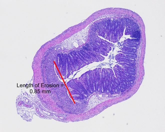
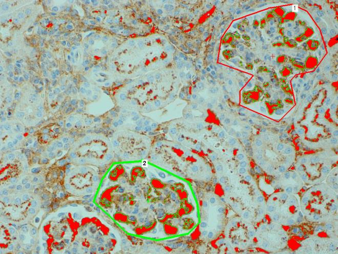
Quantitative Image AnalysisMore advanced image analysis using computer programs to quantify information on the slide.
Quantitative image analysis utilizes software to generate measurements from a tissue section. This process is guided by a veterinary pathologist. The process involves several steps but in its most basic form involves acquiring an image from a microscope, importing that image into a software program, and utilizing tools in that program to process the image and generate data. Whole tissue slide scans can also be analyzed in this way. Some examples include: 1) length or area of specific lesions or features; 2) number of cells stained with a particular marker; 3) amount of one type of tissue identified with a special stain; or 4) cytoplasmic, membrane or nuclear staining of immunohistochemical targets.
|
|
|
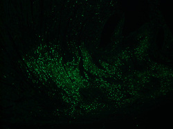
Fluorescent MicroscopyUse of fluorescent labels and antibodies to recognize specific cell types
Use of fluorescent markers can allow for detailed examination of drug uptake, antibody co-localization, or use of antibodies not well suited for brightfield microscopy. We can view fluorescently stained slides and analyze them in the same way as brightfield images, as described above. Our veterinary pathologists have extensive experience with fluorescent microscopy.
|
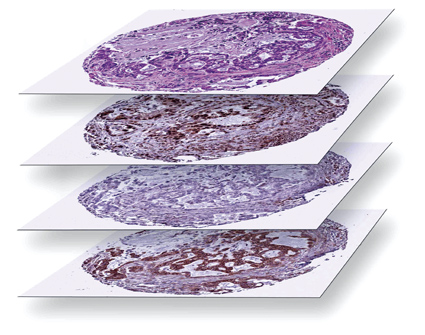
Digital PathologyDigitized, scanned whole slides allow for more advanced image analysis
Whole slides are scanned at high resolution and viewed on a high resolution screen where they can be assessed visually by our pathologists. Because of the extremely high resolution of the scans, advanced computer programs can perform detailed image analysis across an entire study or group of slides with little variability.
|
|